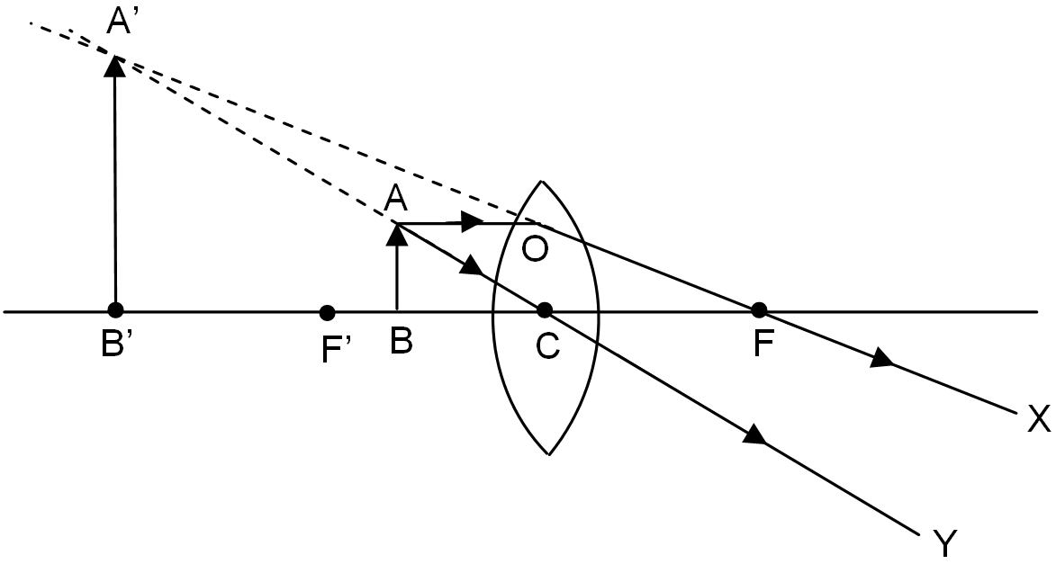
39 compound microscope ray diagram
While JSM-6701F cold field emission scanning electron microscopy (SEM-EDS) was employed to observe the microstructure and fracture morphology of the samples, D/MAX-2400 X-ray diffractometer (XRD) was utilized to analyze the cross-sections at each covering plate temperature, having a step width of 0.02° and stay of 0.12 s at each step. Sodium chloride / ˌ s oʊ d i ə m ˈ k l ɔːr aɪ d /, commonly known as salt (although sea salt also contains other chemical salts), is an ionic compound with the chemical formula NaCl, representing a 1:1 ratio of sodium and chloride ions. With molar masses of 22.99 and 35.45 g/mol respectively, 100 g of NaCl contains 39.34 g Na and 60.66 g Cl. Sodium chloride is the salt …
Oct 13, 2021 · The mass of the coloured compound in the same solution as in question 1 above that would have an absorbance of 1.5 in the same 1.00 cm cell is (in grams): a. 0.050 b. 0.075 log10 2 c. 0.033 log10 2

Compound microscope ray diagram
Nov 07, 2021 · Brightfield Light Microscope (Compound light microscope) This is the most basic optical Microscope used in microbiology laboratories which produces a dark image against a bright background. Made up of two lenses, it is widely used to view plant and animal cell organelles including some parasites such as Paramecium after staining with basic stains. The course of rays through a compound microscopic is shown in the given figure. Here we can see that an image is formed at the least distance of distinct vision ...1 answer · Top answer: Hint: A compound microscope is an optical instrument used for observing highly magnified images of tiny objects. The compound microscope has two lenses ... Friction stir lap welding (FSLW) is expected to join the hybrid structure of aluminum alloy and steel. In this study, the Al-Mg-Si aluminum alloy and 301L stainless steel were diffusion bonded by FSLDW with the addition of 0.1 mm thick pure Zn interlayer, when the tool pin did not penetrate the upper aluminum sheet. The characteristics of lap interface and mechanical properties of the joint ...
Compound microscope ray diagram. Oct 13, 2021 · With the hemispherical shaped biomimetic compound eye demonstrated in Fig. 3e, the identical incident angle of 180° along both the x … Thus, a ray diagram is used to find the final image distance and to determine the total magnification. The overall magnification of a compound microscope is ...3 pages X-ray diffractometer (Rigaku, Japan, D/Max-2400 type, Cu Ka radiation, λ = 1.54056 Å, scanning range of 10-90 º, step length of 0.02 º) was used to analyze the phase structure of the samples. The microscopic morphology and particle size of the materials were observed by scanning electron microscope (SEM, JSM-6701F) and high-resolution ... Scanning electron microscope (SEM) and Energy Dispersive X-Ray Spectroscopy (EDS) were used to evaluate surface morphologies and acquire compositional information of the textured glass at ...
Dec 11, 2020 — I am trying to draw the ray diagram of a compound microscope. Here is my attempt (sorry if the image is kind of blurry):.1 answer · Top answer: Consider an object at the location of the intermediate image between the lenses and consider two rays in particular directions from that object (even ... The history of the telescope can be traced to before the invention of the earliest known telescope, which appeared in 1608 in the Netherlands, when a patent was submitted by Hans Lippershey, an eyeglass maker. Although Lippershey did not receive his patent, news of the invention soon spread across Europe. The design of these early refracting telescopes … Synthesis of BiVO 4 (BVO). Firstly, 3 mmol Bi(NO 3) 3 ·5H 2 O (1.4553 g) and 3 mmol NH 4 VO 3 (0.3510 g) were dissolved in 20 mL ethylene glycol and distilled water respectively and stirred for half of an hour, respectively. When the two substances were dissolved, the two solutions were mixed, and ammonia water was added to adjust the pH as about 10. Oct 18, 2021 · With that said, a compound microscope is capable of a total magnification ranging from forty times the normal size of the sample to up to 1,000 times ((10x) * (100x)) its normal size. Lesson Summary
Simple Microscope. Compound Microscope. 1. Simple Microscope comprises a single biconvex lens used as a magnifying glass. Compound Microscope comprises 2 or more convex lenses where one lens is an eyepiece and the other one is the objective lens. 2. Natural light is the source to see the object. An illuminator is a source to see the object. 3. Consequently, when it is held close to the eye magnified, an erect and virtual image is formed. On the other hand, a compound microscope has two converging lenses, an eyepiece with moderate focal length and large aperture, objective lens of small focal length and short aperture. Telescope. This device is used to observe objects which are far away. According to the Fe-Al phase diagram, the intermetallic phases with Fe x Al y stoichiometries include the Al-rich FeAl 2, Fe 2 Al 5, and FeAl 3 and the Fe-rich Fe 3 Al and FeAl. 29 29. J. J. Yang, Y. Lu, and H. Zhang, " Microstructure and mechanical properties of pulsed laser welded Al / steel dissimilar joint ," Trans. Nonferrous Met. Soc ... Friction stir lap welding (FSLW) is expected to join the hybrid structure of aluminum alloy and steel. In this study, the Al-Mg-Si aluminum alloy and 301L stainless steel were diffusion bonded by FSLDW with the addition of 0.1 mm thick pure Zn interlayer, when the tool pin did not penetrate the upper aluminum sheet. The characteristics of lap interface and mechanical properties of the joint ...
Enormous stingrays being hand fed by two scuba divers at the Cairns aquarium.
The course of rays through a compound microscopic is shown in the given figure. Here we can see that an image is formed at the least distance of distinct vision ...1 answer · Top answer: Hint: A compound microscope is an optical instrument used for observing highly magnified images of tiny objects. The compound microscope has two lenses ...

Fangruida: New coronavirus research 2020v4.0 electronic version (Davis.k)
Nov 07, 2021 · Brightfield Light Microscope (Compound light microscope) This is the most basic optical Microscope used in microbiology laboratories which produces a dark image against a bright background. Made up of two lenses, it is widely used to view plant and animal cell organelles including some parasites such as Paramecium after staining with basic stains.

Image from page 424 of "Appleton's dictionary of machines, mechanics, engine-work, and engineering" (1861)

Image from page 1042 of "The microscope and its revelations" (1901)
To mix the stool and the chemicals together, this Centers for Disease Control and Prevention (CDC) scientist was shown adding a stool sample to the cell culture medium, along with glass beads, which will suspending the stool in the solution. Then the scientist will shake the test tube, in order to get the stool to come off the glass beads, and disperse throughout the liquid.

Spring aerial beauty

Image from page 20 of "The microscope; an introduction to microscopic methods and to histology" (1901)
Robert Charles Gallo, former Biomedical Researcher. He is best known for his work with the Human Immunodeficiency Virus (HIV), the infectious agent responsible for the Acquired Immune Deficiency Syndrome (AIDS). He was the former Chief of Laboratory of Tumor Cell Biology at the National Institutes of Health. 1980

Image from page 112 of "Light, a textbook for students who have had one year of physics" (1921)

Image from page 305 of "A treatise on physiology and hygiene for educational institutions and general readers .." (1884)

Image from page 19 of "The microscope; an introduction to microscopic methods and to histology" (1899)
My eyes

Fangruida: New coronavirus research 2020v4.0 electronic version (Davis.k)

how to draw Compound Microscope ray diagram step wise how ...

Image from page 31 of "The microscope : an introduction to microscopic methods and to histology" (1911)

Image from page 105 of "The Bell System technical journal" (1922)
The microscope

Image from page 369 of "Electron microscopy; proceedings of the Stockholm Conference, September, 1956" (1957)

Stata Center

(Fang Ruida) Davis. K Coronavirus pneumonia biomarkers and treatment(方瑞达) Davis。K å† çŠ¶åž‹ç—…æ¯’æ€§è‚ºç‚Žç”Ÿç‰©æ ‡å¿—ç‰©åŠæ²»ç–—防治

Stata Center

Image from page 39 of "The microscope and microscopical methods" (1896)

Köhler Illumination - Microscopist.co.uk

Breathing is a process that allows you to get oxygen and, at the same time, eliminate carbon dioxide through exhalation. Only under special conditions, such as underwater, this last process is visible to the naked eye.

Image from page 23 of "The microscope; an introduction to microscopic methods and to histology" (1899)

Single Speed, Blue Vibes.

Image from page 480 of "Scientific amusements" (1890)

History of Imaging – Part 2

Image from page 797 of "A Manual of botany : being an introduction to the study of the structure, physiology, and classification of plants " (1875)

Binocular Compound Light Microscope Diagram Quiz
X-Ray

Image from page 369 of "Electron microscopy; proceedings of the Stockholm Conference, September, 1956" (1957)
sunglases
Simple microscope | Fun Science

Image from page 273 of "Cytology" (1961)

Whanake, (v) in Te Reo Maori means to move onwards, move upwards. This was taken at Piha, Auckland, New Zealand. Piha Beach is famous for it’s stunning beaches but beauty can be found on a clifftop on the way to the beach from the carpark. This photo was taken during a photo expedition when I was out of a job, so the fact that it was taken on a whim on the way to the beach resonates that “it’s not about the destination, it’s about the journey.†It matters not what stage of life we’re in as long as we keep moving and looking for inspiration.
Samuel Broder, M.D., former Director, National Cancer Institute from 1989 to 1995.

Image from page 299 of "A treatise on physiology and hygiene for educational institutions and general readers .." (1887)

Image from page 106 of "The Bell System technical journal" (1922)


Comments
Post a Comment