42 diagram of synapse
Diagram Of A Synapse. bbc gcse bitesize how synapses work higher the animation below shows a synapse between two neurons the synapse article human biology at the synapse the firing of an action potential in one neuron—the presynaptic or sending neuron—causes the transmission of a signal to another neuron—the postsynaptic or receiving neuron—making the postsynaptic neuron either more or ... Synapse Diagram. Location. The functions of synapses depend on where they are found. The three forms based on their location are: axodendritic – axon of one neuron and the dendrite of the next adjacent neuron. This is the most common form of synapse that dominates the nervous system; axosomatic – axon of one neuron and the cell body of the other neuron; axoaxonic – between two axons ...
Chemical Synapse – Basic Structure. The most common type of neuron synapse is the chemical synapse. At chemical synapses, the presynaptic neuron is separated from the postsynaptic neuron by a narrow (20 nm), water-filled space called the synaptic cleft. Neurotransmitter molecules are used by the presynaptic neuron to send a message across the ...
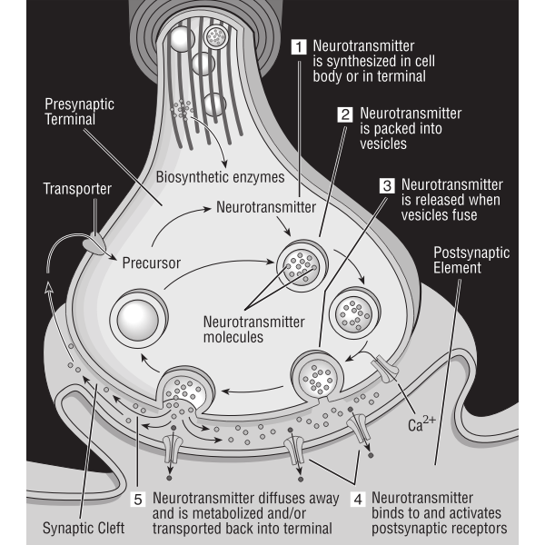
Diagram of synapse
Synapse Diagram Unlabeled. Synaptic Events Worksheet. Use your textbook to complete this activity Label the following parts on the diagram below: Presynaptic neuron Voltage-gated. Download scientific diagram | Example of a synaptic contact between unlabeled cells in the procerebrum. In this and subsequent figures, arrowheads delimit the. The Synapse: The synapse is an area of functional contact between one neuron and another for the purpose of transferring information. Synapses are usually found between the fine terminal branches of the axon of one neuron and the dendrites or cell body of another. This type of neuron is called axo-dendrite synapse. Types. In the central nervous system, a synapse is a small gap at the end of a neuron that allows a signal to pass from one neuron to the next. Synapses are found where nerve cells connect with other nerve cells. Synapses are key to the brain's function, especially when it comes to memory. 1 . The term synapse was first introduced in 1897 by ...
Diagram of synapse. Synapse: Definition, Mechanism and Properties (With Diagram) Article Shared by. ADVERTISEMENTS: In this article we will discuss about:- 1. Definition of Synapse 2. Mechanism of Synaptic Transmission 3. Properties. Definition of Synapse: Synapse can be defined as functional junction between parts of two different neurons. There is no anatomical continuity between two neurons involved in the ... Start studying Synapse. Learn vocabulary, terms, and more with flashcards, games, and other study tools. Chemical Synapse Diagram. chemical synapse at a chemical synapse one neuron releases neurotransmitter molecules into a small space the synaptic cleft that is adjacent to another neuron the neurotransmitters are kept within small sacs called synaptic vesicles and are released into the synaptic cleft by exocytosis the synapse article human biology at the synapse the firing of an action potential ... Types of neurons and synapse structure Each synapse consists of the: Presynaptic membrane - membrane of the terminal bouton (axon ending) of the presynaptic nerve fiber ; Postsynaptic membrane - membrane of the target cell ; Synaptic cleft - a gap between the presynaptic and postsynaptic membranes; Inside the terminal bouton of the presynaptic nerve fiber, numerous vesicles that contain ...
Synapses involving acetylcholine help to modulate signals sent by other neurons. Many neurons can receive chemical messages from several different synapses. As a modulator, acetylcholine may prevent cells from firing and conducting messages, instead of encouraging firing. Activity at the cholinergic synapse, in these cases, provides input that ... About this Quiz. This is an online quiz called Diagram of a Synapse. This quiz has tags. Click on the tags below to find other quizzes on the same subject. Anatomy. Nervous System. Synapse. anatomy 125. Nash-Rule. Synapse - Label. Share Share by Aspillane. Y12 Y13 Psychology. Like. Edit Content. Embed. More. Log in required. Theme. Fonts: Log in required. Options. Leaderboard. Show more Show less . This leaderboard is currently private. Click Share to make it public. This leaderboard has been disabled by the resource owner. ... synapse. 4. The nucleus of a neuron is where genetic material is stored. 5. Neurons that send information from sensory organs, such as the skin or eyes, to the central nervous system are called sensory (or afferent) neurons. 6. Neurons that send information from the central nervous system to muscles or glands are called motor (or efferent ...
The synapse contains a small gap separating neurons. The synapse consists of: a presynaptic ending that contains neurotransmitters, mitochondria and other cell organelles. a postsynaptic ending that contains receptor sites for neurotransmitters. a synaptic cleft or space between the presynaptic and postsynaptic endings. Hear IT!: The synapse image is clearly outlined in the diagram below. [Image will be uploaded soon] Types of Synapse. A synapse can either be a chemical synapse or an electrical synapse depending upon the kind of signals it permits. It's important to understand that even though an electrically excitable neuron generates electrical impulses due to the ... At a synapse, one neuron sends a message to a target neuron—another cell. Most synapses are chemical; these synapses communicate using chemical messengers. Other synapses are electrical; in these synapses, ions flow directly between cells. At a chemical synapse, an action potential triggers the presynaptic neuron to release neurotransmitters. Synapse Labeled Diagram and Receptor. A labeled diagram of a synapse with receptor physiology. Motor neuron, detailed and accurate, labeled. Neuron and Synapse Labeled Diagram. Neuron and Synapse Labeled Diagram. Nerve cells and synapses are implicated in many diseases.
Synapse SQL leverages a scale out architecture to distribute computational processing of data across multiple nodes. Compute is separate from storage, which enables you to scale compute independently of the data in your system. For dedicated SQL pool, the unit of scale is an abstraction of compute power that is known as a data warehouse unit. For serverless SQL pool, being serverless, scaling ...
Diagram of Neuron. A neuron is a type of cell that is largely responsible for conveying information via electrical and chemical impulses. The brain, spinal cord, and peripheral nerves all contain them. The nerve cell is another name for a neuron. The structure of a neuron changes depending on its form and size, as well as its function and location.
Synapses. Where two neurons meet there is a small gap called a synapse. An electrical impulse cannot directly cross the gap so a different mechanism has to be used. An electrical nerve impulse ...
Synapses Where two neurons meet there is a small gap called a synapse . The plasma membranes of each neuron are in very close contact and are separated by a narrow space called a synaptic cleft .
Synapse Color Diagram. High Resolution Poster. Click Here. Citing Research References. When you research information you must cite the reference. Citing for websites is different from citing from books, magazines and periodicals. The style of citing shown here is from the MLA Style Citations (Modern Language Association).
The blink reflex involves synapses. Below is a diagram of a synapse. Identify A, B and C. A _____ B _____ C _____ (Total 3 marks) 4. www.examqa.com Page 6 of 28. The blink reflex can be affected by anaesthetics. Local anaesthetics are used to stop people feeling pain but do not make them unconscious. ...
Calcium channels and the cytoskeleton at the immunological synapse. a Zoomed-in schematic diagram of region shown by box in the lower right panel of Fig. 2a, showing T-cell molecules concentrated at the synapse, separated by a narrow synaptic cleft (∼15-20 nm) from the B cell. Scales are approximate.
Figure 7-2 is a diagram of a synapse. (A) Identify by coloring the following structures, which are typically part of a chemical synapse. (B) Bracket the synaptic cleft Identify the arrows showing (1) the direction of the presynaptic impulse and (2) the direction of net neurotransmitter movements.
What is a synapse? The word synapse stems from the Greek words "syn" (together) and "haptein" (to clasp). This might make you think that a synapse is where brain cells touch or fasten together, but that isn't quite right. The synapse, rather, is that small pocket of space between two cells, where they can pass messages to communicate.
Synapse diagram. Each neuron forms about 2,000 synapses. There are about 10 11 neurons in the CNS. The synapses are of different types and can be classified on the following bases. Parts of neurons involved in the synapse. Axodendritic synapse- The axon of the presynaptic neuron connects to the dendrite of the postsynaptic neuron. This is the most common synapse in the CNS. Axosomatic synapse ...
201 Synapse Diagram Photos and Premium High Res Pictures - Getty Images. United States. CONTENT. Royalty-free Creative Video Editorial Archive Custom Content Creative Collections. SOLUTIONS. Overview Plans and pricing Premium Access Assignments. TOOLS & SERVICES.
Types. In the central nervous system, a synapse is a small gap at the end of a neuron that allows a signal to pass from one neuron to the next. Synapses are found where nerve cells connect with other nerve cells. Synapses are key to the brain's function, especially when it comes to memory. 1 . The term synapse was first introduced in 1897 by ...
The Synapse: The synapse is an area of functional contact between one neuron and another for the purpose of transferring information. Synapses are usually found between the fine terminal branches of the axon of one neuron and the dendrites or cell body of another. This type of neuron is called axo-dendrite synapse.
Synapse Diagram Unlabeled. Synaptic Events Worksheet. Use your textbook to complete this activity Label the following parts on the diagram below: Presynaptic neuron Voltage-gated. Download scientific diagram | Example of a synaptic contact between unlabeled cells in the procerebrum. In this and subsequent figures, arrowheads delimit the.


Comments
Post a Comment