39 amoeba diagram labeled and functions
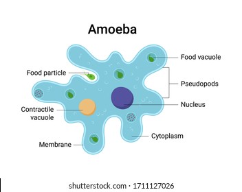
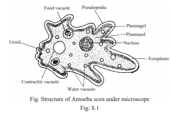
1. Ingestion: The process of ingestion is nothing but intake of food into the body. Amoeba is unicellular and hence it does not have mouth, Amoeba takes the food into the body by forming structures called pseudopodia around the food particle. Structure of amoeba primarily encompasses 3 parts - the cytoplasm, plasma membrane and the nucleus. The cytoplasm can be differentiated into 2 layers - the outer ectoplasm and the inner endoplasm. The plasma membrane is a very thin, double-layered membrane composed of protein and lipid molecules.
Amoeba (plural amoebas/amoebae) is a group of primitive protists. Among the big family of Amoebas, Amoeba proteus is probably the best-known member - common in classrooms and research laboratories. Amoeba proteus is known for the way they move, a primitive crawling manner - through extension and retraction of "false feet" (or pseudopods) over varied substrates.
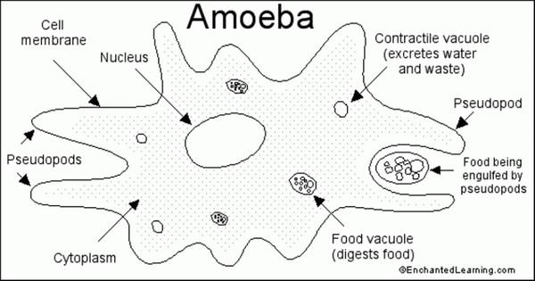
Amoeba diagram labeled and functions
These protozoa typically lack a fixed external anatomy and are flexible but can be categorized by the shape and number of the pseudopodia. Enclosed by this cell membrane (also known as the plasma membrane) are the cell's constituents, often large, water-soluble, highly charged molecules such as proteins, nucleic acids, carbohydrates, and substances involved in cellular metabolism. In amoeba, several digestive enzymes react on the food present in the food Draw a diagram to show the nutrition in Amoeba and label the parts used for this purpose. Digestion in Amoeba. itzaredpanda4 itzaredpanda4 The process shown is the the digestion process in amoeba which consist of absorption, assimilation, and egestion An amoeba (/ ə ˈ m iː b ə /; less commonly spelled ameba or amœba; plural am(o)ebas or am(o)ebae / ə ˈ m iː b i /), often called an amoeboid, is a type of cell or unicellular organism which has the ability to alter its shape, primarily by extending and retracting pseudopods. Amoebae do not form a single taxonomic group; instead, they are found in every major lineage of eukaryotic ...
Amoeba diagram labeled and functions. Amoeba exhibits movement by the pseudopodia. It also helps in food capture. Like an ordinary cell the body of amoeba has 3 main parts: Plasma lemma or plasma membrane, Cytoplasm and nucleus. Plasma lemma is a very thin, delicate and elastic cell membrane of amoeba. It is composed of a double layer of lipid and protein molecules. An amoeba is unicellular and moves by using pseudopods. A pseudopod is a temporary bulge that forms in the cell membrane as a result of the movement of the cytoplasm. The word pseduopod means "false foot." The pseudopod has two functions, or uses: 1. to move, 2. to capture food. The picture below shows an example of a pseudopod in an amoeba. Label Amoeba Diagram Using the definitions listed below, label the amoeba. ... of the amoeba, located centrally; it controls reproduction (it contains the chromosomes) and many other important functions (including eating and growth). pseudopods - temporary "feet" that the amoeba uses to move around and to engulf food. Enchanted Learning Search. Learn more about the anatomy of amoeba using these amoeba diagrams available in clear figures! A total of five diagrams are provided to show you the structure of amoeba. The following anatomy diagrams are available to help you understand more about the anatomy of amoeba. We'll start by giving you the first labeled diagram below.
Amoeba is an aquatic, single-cell (unicellular) organism with membrane-bound (eukaryotic) organelles that has no definite shape. It is capable of movement. When seen under a microscope, the cell looks like a tiny blob of colorless jelly with a dark speck inside it. Some parasitic amoebae living inside animal bodies, including humans, can cause various intestinal disorders such as diarrhea ... Structure and Function. Since Euglena is a eukaryotic unicellular organism, it contains the major organelles found in more complex life. This protist is both an autotroph, meaning it can carry out photosynthesis and make its own food like plants, as well as a heteroptoph, meaning it can also capture and ingest its food. Cell Membrane Of Algae.In most algal cells there is only a single nucleus, although some cells are multinucleate. The eukaryotic algal protoplast is surrounded by a lipoproteinaceous external boundary, called cell membrane, and consists of one or more usually spherical or ellipsoidal nucleus and cytoplasm. Structure and Functions of Amoeba Parts. Cytoplasm - The cytoplasm is differentiated into Ectoplasm and endoplasm. The ectoplasm forms the outer and relatively firm layer lying just beneath the plasma lemma. It is a thin, clear (non-granular) and hyaline layer It is thickened into a hyaline cap at the advancing end at the tips of ...
The amoeba belongs to the kingdom Protista. It is a single celled microscopic organism (about 0.3 mm across). It has cytoplasm, nucleus, cell membrane and a variety of inclusions in the cytoplasm and exhibits all the essential functions of any living organism.They are found in fresh water (like puddle and ponds), salty water, wet soil and in ... Label diagram of amoeba. Proteus and part of what makes the organism so fascinating. Plural amoebas or amoebae ə ˈ m iː b i often called an amoeboid is a type of cell or unicellular organism which has the ability to alter its shape primarily by extending and retracting pseudopods. An amoeba ə ˈ m iː b ə. It helps the amoeba move feed ... Culture of Amoeba proteus. It may be obtained for class study by scraping decaying vegetation from the bottom of a pond. Then scrapping, is settle down in the wide-mouth container, Amoeba of different kinds may be found in the sediment and sorted with the help of fine pipette under a binocular microscope. A temporary culture of Amoeba can be prepared in the laboratory by the hay-infusion method. Structure of Amoeba Proteus - Amoeba proteus is a single-celled organism found in ponds, lakes, freshwater pools, and slow-moving streams. It is a unicellular organism and measures about 250 to 600 µm in maximum diameter and so transparent that is invisible to naked eyes. It usually, feeding on algae, bacteria, and other microorganisms.
Structure of Amoeba. Its structure is studied with the help of a microscope. Its diameter is from 0.25 to 0.60 mm. (1/100 inch). The shape of its body is not definite because pseudopodia are continuously being formed in different directions and thus, the shape of the body keeps on changing.
This will also help you to draw the structure and diagram of amoeba. 1. Fresh water and free living organism commonly available in stagnant water. 2. Body irregular and cytoplasm clearly differentiated into ectoplasm and endoplasm. 3. Body naked, and extends into numerous finger like projections the pseudopodia. 4.
The adult trophic form of Entamoeba is known as Trophozoite or Magna. It inhabits anterior part of large intestine, i.e. colon of human beings. It resembles amoeba in structure but differs in parasitic mode of life. Its body is covered by plasma lemma and cytoplasm is differentiated into ectoplasm and endoplasm.
Articles and drawings on Protoctista, Protista, Amoeba, Paramecium, Spirogyra, Chlamydomonas, Euglena, Malaria, Resources for Biology Education by D G Mackean
Amoeba Anatomy Amoebas are simple in form consisting of cytoplasm surrounded by a cell membrane . The outer portion of the cytoplasm ( ectoplasm ) is clear and gel-like, while the inner portion of the cytoplasm (endoplasm) is granular and contains organelles , such as a nuclei , mitochondria , and vacuoles .
Nutrition in an Amoeba occurs through a process called phagocytosis where the entire organism pretty much engulfs the food it plans on eating up. The mode of nutrition in amoeba is known as holozoic nutrition. It involves the ingestion, digestion and egestion of food material. Amoeba does not have any specialized organ for nutrition.
Amoeba plural amoebasamoebae is a genus that belongs to kingdom protozoa. Labeled amoeba proteus microscope.An electron microscope study of the nuclear membrane of amoeba proteus by thin sectioning techniques has revealed an ultrastructure in the outer layer of the membrane that is homologous to the pores and annuli observed in the nuclear membranes of many other cell types studied by these ...
Amoeba Labeled And Functions Aflam Neeeak. Amoeba Biological Labeled Vector Illustration Diagram. Biology Diagram Of Paramecium And Amoeba With Their. ... Labeled Amoeba Diagram By Sciencedoodles On Deviantart. Amoeba Drawing At Getdrawings Free Download. Amoeba Drawing At Explore Collection.
Amoeba. Body structure: Amoeba appears as a colourless and transparent drop of jellywhen viewed under a microscope. It lacks a definite body shape because it changes its shape by producing the pseudopodia every moment. So, it is not possible to describe its definite shape, anterior or posterior ends, dorsal and ventral surfaces.
Biology Protista Amoeba Malaria Paramecium Spirogyra Chlamydomonas Euglena Educational Notes Drawings By D G Mackean
Pseudopodia - Structure and Function: A labelled diagram of Amoeba proteus can be seen above.The pseudopodia are the most defined structures of A. proteus and part of what makes the organism so fascinating.These "false feet" are used for movement and to engulf prey (see Nutrition for further detail) - making it an essential part of its structure.
An amoeba is a unicellular, highly motile eukaryotic organism that can be found in its free-living form in various water bodies and as a parasite in both vertebrates and invertebrates.The organism belongs to the kingdom protozoa. The general scientific name of amoeba is Amoeba proteus. There is some parasitic amoeba such as the intestinal parasite Entamoeba histolytica and the brain-eating ...
An amoeba (/ ə ˈ m iː b ə /; less commonly spelled ameba or amœba; plural am(o)ebas or am(o)ebae / ə ˈ m iː b i /), often called an amoeboid, is a type of cell or unicellular organism which has the ability to alter its shape, primarily by extending and retracting pseudopods. Amoebae do not form a single taxonomic group; instead, they are found in every major lineage of eukaryotic ...
In amoeba, several digestive enzymes react on the food present in the food Draw a diagram to show the nutrition in Amoeba and label the parts used for this purpose. Digestion in Amoeba. itzaredpanda4 itzaredpanda4 The process shown is the the digestion process in amoeba which consist of absorption, assimilation, and egestion
These protozoa typically lack a fixed external anatomy and are flexible but can be categorized by the shape and number of the pseudopodia. Enclosed by this cell membrane (also known as the plasma membrane) are the cell's constituents, often large, water-soluble, highly charged molecules such as proteins, nucleic acids, carbohydrates, and substances involved in cellular metabolism.

Amoeba Amoeba Diagram Amoeba Image Labelled Diagram Amoeba Amoeba Labelled Diagram Science Diagram Youtube


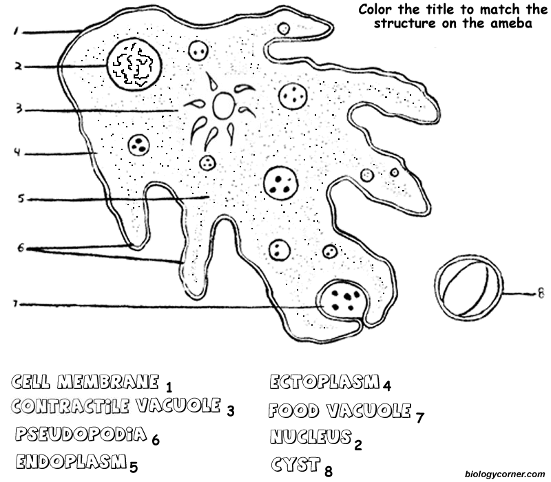


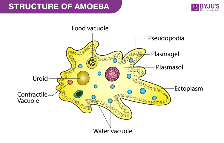

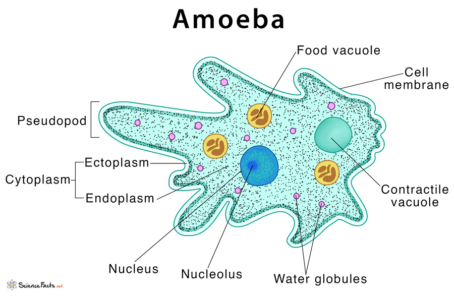





Comments
Post a Comment